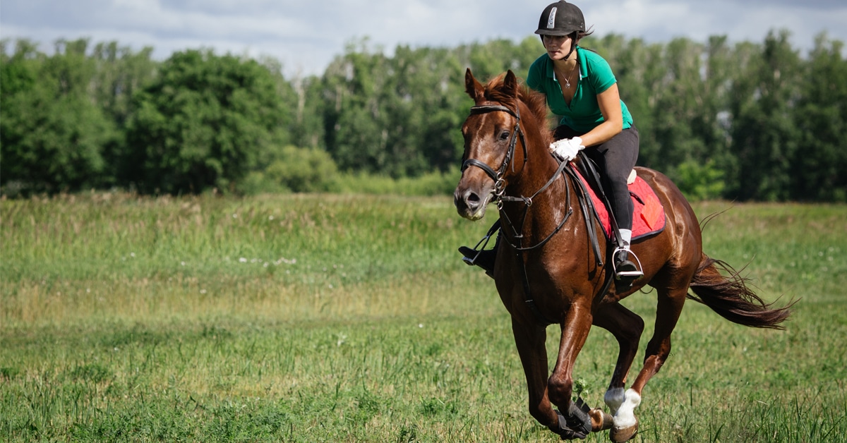
Laminitis is caused by a disruption in the junction between the sensitive and insensitive tissues of the hoof, known as the basement membrane. This leads to a loss of structural support between the hoof wall and the distal phalanx (or coffin bone), the last bone in the foot. The bone then rotates or sinks within the hoof.
Once bone rotation happens, affected horses are more prone to future incidents of laminitis because the hoof wall-bone connection never re-forms satisfactorily. In some cases, the disease can progress to the point that the bone comes through the sole of the foot, which typically requires euthanasia of the horse for humane reasons.
3 Phases of Laminitis
Developmental – when the triggers that lead to laminitis are at work. This stage lasts about 20 to 60 hours after exposure. Clinical signs are not noticeable.
Acute – pain and lameness become evident as the laminae become inflamed. Indications of laminitis include: short, choppy stride; a sawhorse stance (front feet forward, rocked back on their hind feet); shifting weight from side to side; an increased (frequently called “bounding”) digital pulse; heat in the hoof; and pain when pressure is put on the toe with hoof testers. The horse may also appear anxious, stop eating and show an increased respiration rate. This phase lasts 24 to 72 hours.
Chronic – when clinical signs have been present for 72 hours or when x-rays show coffin bone rotation. Some experts call this stage the sub-acute phase, and consider the condition chronic when permanent damage has occurred. Signs of long-term laminitis problems include: rings in the hoof wall; bruised soles; seedy toe; dropped soles; dished hooves; toe cracks; and flakey outer hoof wall.
— Nicole Kitchener
What causes laminitis?
Laminitis is a poorly understood disease with many causes. These reasons range from primary events such as ponies grazing on lush pasture or grain-overloaded horses, to secondary events such as systemic diseases.
For example, horses that have been diagnosed with metritis (uterine infection), colitis (diarrhea) or metabolic conditions such as pituitary pars intermedia dysfunction (PPID) can be prone to laminitis. As well, horses that have undergone orthopedic surgery or are recovering from an existing lameness may develop supporting-limb laminitis.
In primary events such as grain overload, researchers believe the disease is a result of the overgrowth of gram-positive bacteria (Streptococcus species) in the hindgut. As the bacteria overgrow and the gut becomes inflamed, the barrier between the gastrointestinal tract and the blood becomes more permeable. Bacteria and their by-products more easily enter into the horse’s systemic blood circulation.
These bacteria produce both endotoxins and exotoxins (see more on page 23). When they enter the horse’s bloodstream, these products give rise to signs of systemic illness such as fever and elevated white blood cell counts. Once the horse recovers from the initial illness, they go on to develop laminitis – presumably because of the by-products of endotoxemia and local inflammatory changes.
In secondary events where laminitis occurs after the horse suffers from a systemic disease, there is a body-wide inflammatory disease process to which the hoof’s laminar tissues are very sensitive.
Laminitis as a result of metabolic conditions such as PPID is even less understood. It may be that horses with PPID have an underlying problem with the structure of the hoof due to the primary endocrine disorder. The hoof is then at increased risk simply because it’s less structurally sound to begin with.
There is also some evidence that some horses may have a genetic predisposition to laminitis, but this theory hasn’t been proven yet.
How is laminitis treated?
Traditionally, laminitis is treated in the acute stages with non-steroidal anti-inflammatory drugs (NSAIDs). The most common NSAIDs used by veterinarians are phenylbutazone (Bute) or flunixin (Banamine®). These medications provide pain relief and dampen the inflammatory response. In my experience, phenylbutazone is more effective in treating the condition.
Other treatments include icing the horse’s feet in the attempt to reduce local inflammation in the affected hoof. While this practice seems a little far-fetched, reducing the temperature of horse’s feet has several important effects:
- Icing an inflamed region makes it feel better as it numbs the pain.
- In horses with laminitis, some of the proposed toxins may be temperature-sensitive and less damage occurs at lower temperatures.
- Icing the feet is often not enough. Researchers in Australia have shown that cooling the entire limb from the knee (carpus) and hock down is necessary to significantly reduce foot temperature in a horse.
Commercial cooling products are similar to those used by human athletes for sports injuries. However, researchers at the Western College of Veterinary Medicine (WCVM) found that these products provided insufficient cooling in a clinical trial situation. Icing the feet with bags and frequently changing the ice (or having the horse stand in an ice bath) was the most successful in reducing the clinical signs of the disease.
External structural support systems such a supportive shoeing, foam pads and soft-ride boots are useful therapeutic options that veterinarians often use in combination with deep bedding to cushion the foot and provide counter-pressure to the bone rotation. Some horses choose to lie down, which will also takes the pressure off their feet and reduce pain.
Once laminitis becomes a chronic condition, a good farrier becomes essential. Through regular trimming of the hoof, the farrier will attempt to regain as close to normal weight-bearing angles and forces that act on the foot. Radiographs (x-rays) to follow the progression of disease and to monitor the long-term health of the foot can be extremely useful.
Can laminitis be prevented?
Strict diet control and nutritional management can help in the cases of ponies or horses that have previously foundered. Keeping the horses away from grain bins or lush grass will also help.
Proper weight management is essential. There is some evidence that overweight horses can develop insulin resistance – leaving them more prone to laminitis.
Treatment of underlying metabolic conditions, such as properly-managed PPID, can go a long way to reducing the number of recurring bouts of laminitis. Affected horses should be provided with medical management and veterinary care.
Laminitis Research
Many researchers around the world are working to understand the disease process of laminitis. By gaining a better understanding of the condition, we can better manage or prevent the disease.
A few examples of laminitis research studies include looking at the vascular changes that occur within the hoof, studying the activation of local inflammatory molecules and investigating the hoof’s thermodynamics as an indicator of the onset of a laminitic episode.
Throughout North America, there is an ongoing laminitis surveillance program through the American Association of Equine Practitioners (AAEP). Based out of Texas A&M University, The Laminitis Project is a prospective epidemiological study that’s looking at new acute cases of pasture-induced and metabolic laminitis cases. It’s the first large-scale study of this kind and gathering this information from horse owners and veterinarians may help to better identify at-risk horses. The results are not yet available, but you can learn more at vetmed.tamu.edu/laminitis.
At the WCVM, there is research being done to develop a vaccine for laminitis. This vaccine would be effective in cases that occur as a result of a systemic inflammatory response to the hindgut overgrowth. It targets the exotoxin produced by the Gram-positive bacteria.
So far, the vaccine has been shown to be protective if given before development of laminitis. Given as a preventive measure, vaccination may provide protection against laminitis – and hope for horses
in the future.
Endotoxins vs. Exotoxins
Endotoxins – Associated with the outer membranes of certain gram-negative bacteria. Endotoxins are not secreted, but are released only when the cells are disrupted; they are less potent and less specific than exotoxins. In large quantities they produce hemorrhagic shock and severe diarrhea; smaller amounts cause fever, altered resistance to bacterial infection, leukopenia followed by leukocytosis, and numerous other biologic effects.
Exotoxin – A toxin secreted by bacteria. An exotoxin can cause damage to the host by destroying cells or disrupting normal cellular metabolism. They are highly potent and can cause major damage to the host. Exotoxins may be secreted, or, similar to endotoxins, may be released during lysis of the cell.
The Latest