Healing a wound is a hot topic at our Canadian veterinary schools. Several interesting studies are currently being conducted at the University of Calgary Faculty of Veterinary Medicine (UCVM), the Western College of Veterinary Medicine (WCVM) and the Université de Montréal Faculté de Médecine Vétérinaire (FMV).
Improving Grafting
Dr. Holly Sparks of the UCVM is researching the use of stem cell-based therapies to improve skin grafting techniques for horses and humans with traumatic skin wounds and burns.
Dr. Sparks received the prestigious Alberta Innovates: Health Solutions Clinician Fellowship Award to fund her research over the next three years. She will study the use of dermal (skin) stem cells as well as novel bioengineered ‘scaffolds’ to place new, healthy cells on top of wounds to regenerate and promote healing.
Currently, the gold standard of burn or large wound treatment is ‘split thickness’ skin grafting, where the top layer of skin is harvested from a healthy area and is placed onto the wound to close it. The problem, Dr. Sparks explained, is the grafted skin lacks the full complement of the supporting middle layer of skin that includes hair follicles and glands, so it’s prone to scarring, itchiness and pain. “Our aim is ultimately to regenerate that lost dermis and improve the function of skin grafts. To do that, we’re culturing and implanting dermal stem cells underneath the grafted layer of skin. Previous work has shown these cells can regenerate new dermal tissue in mice and stimulate new hair follicle formation.”
Dr. Sparks is working with colleagues in engineering to create a bioengineered scaffold – a microscopic sheet of woven material to more easily deliver cells into wounds, and allow them to survive and begin generating new tissue. “It’s a huge project and very collaborative. Our team is comprised of not only basic scientists, engineers and veterinarians, but also human physicians specializing in burn wounds and plastic surgery.”
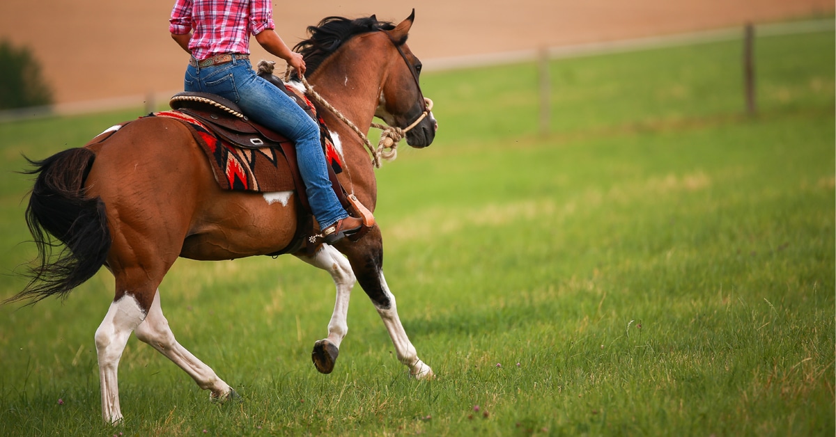
Pony Power
Meanwhile, researchers at the WCVM are using stem cells in a different way to improve wound healing. According to studies, skin wounds in horses heal slower than in ponies, whose initial inflammatory response to wounds is much greater. As a result, ponies’ wounds heal faster and their leg wounds don’t develop proud flesh (an excessive growth of granulation tissue), which is a common problem in horses. Dr. Spencer Barber and a team of researchers believe that if the inflammatory response of an adult horse can be manipulated to mimic that of a pony, it will increase the rate of healing in that horse. Their theory is based on previous WCVM studies which investigated the behaviour of equine cord blood stem cells once they were injected into the jugular veins of horses participating in the research. Based on the team’s preliminary analysis of skin wound biopsies from the animals, it appears that injected stem cells do migrate to wounds.
In this prospective study, the researchers will continue their analysis of the wound biopsy tissues collected in their clinical study. Other researchers have already proven that stem cells promote healing in laboratory animals, but the WCVM team’s stem cell work with live horses is the first of its kind. Once their analysis is complete, they hope to determine whether injecting stem cells from ponies’ umbilical cords can positively influence the quality of wound repair and speed up the healing process in horses’ wounds.
Oxygen Needed
Dr. Christine Théorêt has been participating in an ongoing study at the FMV investigating the link between hypoxia (low levels of oxygen) and the formation of proud flesh on limb wounds in horses, as well as the genes that play a role in its development.
Studies have shown that hypoxia in living tissue stimulates the growth of new blood vessels and granulation tissue, which is a normal part of the wound healing process known as angiogenesis. Dr. Théorêt’s team has discovered that healing equine limb wounds are deficient in anti‐angiogenic molecules relative to body wounds that do not develop proud flesh, suggesting that the “control switch” to limit blood vessel formation is defective in limb wounds.
In particular, they noticed that the degree of oxygen saturation in limb wounds is inferior to that of body wounds during the early period of healing, indicating a temporary, relative state of hypoxia during the inflammatory phase of repair. They also observed that blood flow is significantly reduced in the new blood vessels that form in limb wounds compared to those in body wounds.
Ongoing studies in mice are hoped to provide answers on increasing oxygen and/or circulation to limb wounds.
The Latest