On November 6, 2011, Hickstead collapsed shortly after he and Eric Lamaze completed their first round at the Rolex Grand Prix of Verona CSI in Italy. Despite instant medical attention from several veterinarians, the 15-year-old Dutch warmblood stallion would never again rise to his feet. After a necropsy was performed, it was determined that Hickstead had succumbed to an aortic aneurysm. The physical capabilities of the equine heart are extraordinary. A horse’s ability to amplify his heart rate during exercise to nearly 10 times greater than his resting rate is a key reason for his athletic prowess.
But functional or structural abnormalities can occur. “Next to respiratory and musculoskeletal disorders, cardiac disease represents the third most important cause of loss of performance in horses,” says Dr. Robert Gilmour, DVM, of Cornell University in Ithaca, NY.
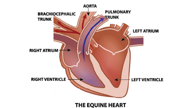
A powerful pumping machine
A horse’s heart is roughly the size of a large melon and consists of the left and right receiving chambers; left and right pumping ventricles; left and right inflow valves and outflow valves; and attached inflow veins and outflow arteries. The left side of the heart gathers oxygenated blood returning from the lungs and pumps it to the body through the aorta. The right side collects blood filled with cellular waste from the body and delivers it to the lungs through the pulmonary artery.
A heart beat consists of a contraction/ejection phase, or systole, and a relaxation/filling phase, or diastole. The heart is triggered to contract by electrical cues from its muscles.
Stroke volume is the amount of blood pumped in each beat and in an adult horse at rest can range from 25-40 liters per minute. Horses, however, can increase their stroke volume up to 50 percent through training.
Like other muscles, the heart muscle remodels in response to exercise and can increase in size. Endurance training increases the overall size of the heart’s left chambers, while sprint training increases the thickness of the ventricle walls. These adaptations enable the heart to pump greater quantities of oxygenated blood more efficiently.
“Athletic training in horses results in profound effects on cardiac morphology,” says Dr. Lesley E. Young, BVSc, PhD, of the Center of Equine Studies of Newmarket, UK. “Sports specific adaptations to exercise occur in the horse, as in other athletic species, and we have recently shown that these responses are appropriate to the endurance component of the horse’s primary athletic discipline. The average increases in relative cardiac dimensions in horses subjected to training are large, ranging from 10 to 33 per cent.”
The heart, along with the lungs, blood vessels and cell mitochondria, plays a major role in the amount of oxygen a horse is able to process at any given time. Called VO2 max, this amount is derived by simply multiplying the stroke rate by the heart rate for the total cardiac output. A top human athlete can process 80 millilitres of oxygen per kilogram per minute, while a fit horse can consume 180 millilitres of oxygen per kilogram per minute. It is generally accepted that horses have the largest capacity to process oxygen, actually in much greater amounts than the heart and lungs supply, making them pound-for-pound the world’s greatest athletes.
Since the cardiovascular system can be enhanced and pulmonary function cannot, increasing a horse’s total cardiac output augments their ability to perform. “It has been found that maximum oxygen consumption increased by 23 per cent due to increases in cardiac output and stroke volume,” explains Dr. Susan Holcombe, VMD, PhD, College of Veterinary Medicine at Michigan State University. “But pulmonary function was either unchanged or deteriorated. Therefore, it would seem horses would be limited by their respiratory system.”
Diagnostic tools
The two main tools veterinarians use to diagnose equine heart abnormalities are ultrasound, or echocardiogram, and electrocardiogram (ECG). An echocardiogram uses sound waves to penetrate the body’s tissues and provide an image of the heart’s structure by reflected light. The machine then produces a two-dimensional image (sonogram) that tabulates the results. An ECG ascertains the rhythm of the heart by measuring its electrical activity through electrodes placed around the thoracic cavity. Since the horse has a large cardiac reserve, using an ECG at rest provides very limited information; it is during exercise that ECGs are an important tool to evaluate horses.
Heart rate monitors using global positioning system (GPS) technology are widely used in sport and event horses. Wireless electrodes are sewn into a belt that can be worn on its own or placed under a horse’s saddle. Dr. David Evans, BVSc, PhD, associate professor in Veterinary Science at the University of Sydney in New South Wales, AUS, sought a reliable, durable method for trainers to assess their horses’ fitness and deduced GPS would be an excellent option because it can measure velocity and position at frequent time intervals in real training situations.
“Measuring maximum oxygen uptake is the gold standard for aerobic fitness, but it involves putting a mask on the horse’s face and exercising the horse at high speeds on a treadmill,” Dr. Evans says. “Heart rate is another fundamental measurement used during exercise to determine fitness and this method (GPS) is accurate and precise. You can measure directly without observing the horse at all times and it can be used to record sectional times and peak velocities.”
According to Is Your Horse Fit? The Physiology of Conditioning, published by the Alberta Agriculture and Rural Development, measurement of recovery heart rate becomes the best option for assessing the horse’s fitness level. Record the heart rate at 2, 5 and 10 minutes after completion of exercise; in well-conditioned horses, the heart rate will be around 60 bpm after 10 minutes of recovery, even following reasonably strenuous workouts. However, a recovery heart rate of 72 to 80 bpm or more might indicate that the work effort was beyond the horse’s current level of conditioning.
Common abnormalities
Functional or structural abnormalities can seriously alter cardiac output during exercise, or even at rest. The structural soundness of the heart, how strongly the ventricles are able to contract, how easily the ventricles are able to fill during relaxation, and if there is any resistance in the ventricles to normal blood flow are all factors.
Cardiac murmurs are prolonged sounds during periods of the cardiac cycle that are usually silent and arise when normal blood flow is disrupted. “When blood flows smoothly through the heart and blood vessels, very little sound is produced, like water flowing through a hose,” states Dr. William Thomas, DVM, of the University of California Davis Center for Equine Health. “The blood flow becomes turbulent because of increased velocity and you would receive a spraying sound like when you put your thumb across the end of the hose.”
Murmurs usually do not affect a horse’s performance. “Cardiac murmurs occur in 65-80% of athletic horse breeds, including Thoroughbreds,” says Dr. C.M. Marr, BVMS, PhD, of Rossdales Equine Hospital in Newmarket, UK. “While murmurs can be associated with poor performance or even occasionally heart failure, they are usually discovered as part of a general physical examination; for example, in a pre-purchase setting or prior to anaesthesia.”
Congenital defects are abnormalities present at birth and are very rare in horses. The most common equine congenital defect is ventricular septal defect, which is a hole in the wall between the two ventricles. “Congenital defects occur less commonly than the other causes of murmurs and typically are very loud,” says Dr. Marr. “Simple defects, such as the ventricular septal defect, are often well-tolerated and the adverse affect on cardiac function is minimal.”
Atrial fibrillation, or arrhythmia, is an electrical disorder that causes an irregular heartbeat and is more common in the horse than in other domestic species. In adult horses at rest, a regular heart rate of 26-50 beats/minute is considered normal. When a horse exercises, his heart rate typically exceeds 200 beats per minute. During atrial fibrillation, there is chaotic electrical activity which causes a horse’s atria to contract up to 300-400 times per minute. This condition significantly reduces cardiac output, which in turn, profoundly compromises a horse’s performance.
“There is a high incidence of atrial fibrillation in horses because of their large atrial size,” explains Dr. G. van Loon, DVM, PhD, of the Dept. of Large Animal Internal Medicine at Ghent University in Belgium. “In Thoroughbreds, atrial fibrillation has been shown to occur relatively frequently during a race. In these animals, it disappears spontaneously shortly after termination of exercise and may remain undiagnosed if the horse is not immediately examined.”
Sudden death – but not a heart attack?
Although most people consider the cause of death for a horse that suddenly expires to be a heart attack, nearly all these sudden-death situations (which occur in less than 1% of the entire equine population) are actually ruptures. It is very rare for a horse to have heart disease like humans, because their arteries don’t clog from cholesterol buildup, so they would not experience an actual heart attack as we know it.
An aneurysm – an area of a blood vessel (normally an artery) that has become enlarged – is more likely the culprit. When aneurysms rupture, they can cause hemorrhage into adjacent tissues, loss of blood flow and, as in Hickstead’s case, death. This condition can be caused by congenital weakness of the blood vessels, copper deficiency, external trauma or mechanical failure. As there is not enough documentation to completely prove horses are prone to genetic disorders or copper deficiency, it is supposed the predisposing factors for horses are degeneration of the vessels by the aging process and stress-related weakening of the artery walls. As the aorta is subject to high pressures, ruptures in horses usually occur in this vessel.
“When rupture does occur, the consequences are most often immediately fatal, a consequence of blood loss, and when not so, are nonetheless associated with distress (due to pain), and a sudden and severe reduction in performance and readiness to undertake work,” says Dr. Peter Physick-Sheard from the Ontario Veterinary College. “Sudden or progressive development of an aneurysm can be a consequence of a split forming in the wall of an artery, with blood dissecting along tissue layers in the wall of the artery to form a dissecting aneurysm. Affected horses may have rapid and shallow respirations, very high heart rates, possibly some head and neck extension, will move cautiously, and may appear to be in distress. The onset will be sudden, and if the problem occurs during exercise, the horse will stop suddenly and may adopt a base-wide stance with the forelimbs.”
Since most horses will never exhibit any signs of developing an aneurysm prior to its rupture, this condition is nearly impossible to ascertain, as veterinarians do not possess the diagnostic capabilities to pinpoint an aneurysm before it becomes an issue. “Since changes can be developing slowly over many months, and are usually asymptomatic until the acute event takes place, they can be hard to diagnose,” adds Physick-Sheard. “In humans, there are a range of surgical approaches that can be taken to strengthen or even replace the region of damaged artery, but this is neither feasible nor desirable in an animal like the horse that is used for athletic activity. If the horse shows evidence of thoracic pain, an ultrasound examination can be used to identify the lesion, though this may require the use of trans-oesophageal ultrasound. Complete retirement from athletic activity is the only option, and in some cases humane destruction may be advisable if there are potential dangers for persons working around the horse.”